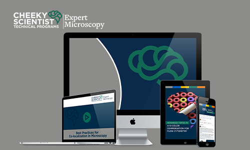
Here's What You Get Immediate Access To When You Become An Exscope Member
The Expert Microscopy Career Program Gives Scientists Like You Certainty So You Can…
- Understand what it takes to capture an image that meets the approval of editorial, grant, and patent reviewers.
- Create images and perform statistical analyses on your images that will impress your supervisor and put you in a better position for a promotion.
- Complete experiments more quickly so they avoid wasting time and resources.
- Lessen the effort involved in capturing important microscopy images.
- Decrease the frustration that results from a poor understanding of microscopy principles.
- Lay a solid foundation for biotech experiments or more advanced research.
- Gain important information that enables them to troubleshoot microscopy challenges during research.
- Follow best practices for avoiding accusations of image fraud.

The Expert Microscopy Career Program
This step-by-step program provides both students and researchers with a crash course in microscopy—giving them a broad, but specific, knowledge in principles that will help them perform their experiments faster and better.
This program is ideal for core managers who need a curriculum for their students, lab managers who want to shore up knowledge gaps, and graduate students or scientists who desire a better foundation in microscopy.
Throughout this course, you’ll find that I’ve communicated important concepts in an understandable way—which means that individuals with a variety of academic backgrounds can grasp this material. (In other words, you don’t need a background in physics to enroll in this course.)
On top of that, this program also provides access to a private group where Expert Microscopy Career Program students can get their questions answered.
No judging. No raised eyebrows. Just helpful support for the challenges students are facing.
Between the modules and the private support group, this program can help you or your team spend less time troubleshooting problems…and more time accomplishing important research goals.

“I have reviewed the ExScope course and found the material to range from basic to complex ideas that are conveyed in a succinct manner. There are consolidated, practical tips for preparation, imaging, processing and post-processing. It is a useful resource for anyone planning to begin working with microscopes which can go on to serve as a reference for advanced users too.”
“This course is a must-have for anyone interested in or looking to expand their knowledge of microscopy. Each module is well-organized and easy-to-follow. The resources provided both reinforce what the user learned and offer more detail about microscopes and microscopy techniques. This course is a one-stop-shop for microscopists of all skill levels! ”
Whether You’re A Lab Manager Looking To Educate Your Students, A PhD Or A Postdoc…
See If You Answer Yes To These Questions:
- Are you embarrassed by subpar images that poorly reflect the quality and effort invested in a research project?
- Are you frustrated because no one ever taught you (or your students) how to properly prepare samples for imaging?
- Are you ready to put a stop to delayed research goals…that can be traced back to a poor understanding of the basics of microscopy?
- Are you tired of knowledge gaps that lead to wasted resources in the lab?
- Are you concerned that important research projects will never achieve the recognition they deserve…simply due to poorly captured images?
- Are you ready to experience more self-sufficiency in the lab…or help your colleagues be more self-sufficient?
- Are you ready to drive science forward with crisp, clear images that introduce scientific discoveries?
Microscopy Plays A Crucial Role In Scientific Experiments, Bringing New Insights To Life.
There’s Nothing Like A Brilliant Image To Underscore A Scientific Breakthrough.
The Expert Microscopy Career Program Mission
Curious how I came to put together a course on microscopy?
My journey in microscopy reaches back to when I was in graduate school—working on homebuilt microscopes. With these devices, if something goes wrong, there’s no one to call. It’s up to you to troubleshoot your problem and find a way forward to continue your research.
My research experiences led me to hone my microscopy skills, and—eventually—I landed a position as an account manager for Zeiss.
From MIT to the UMass System, I was responsible for helping the universities in my care find the right microscope solutions to support their students and research…I even played a role as commercial faculty for Zeiss at Advanced Quantitative Light Microscopy at Woods Hole Marine Laboratory.
Today, I’m a core manager and an assistant director of microscopy at Wake Forest University, where I help students better understand microscopy, help plan experiments that answer their scientific questions precisely, and obtain funding for equipment that will further the university’s research goals.
That’s been my professional journey, but here’s a big reason behind why I put together these modules: I love teaching microscopy.
I’m passionate about helping people capture high-quality microscopy images…revealing something that no one has ever seen before.
In fact, capturing excellent images can play an important role in moving science forward.
Having a visually stunning image of your research specimen can tip the scales in your favor when it comes to getting your work published.
But creating stunning images doesn’t happen by accident.
It takes skill, expertise, and careful execution

Join The Expert Microscopy Career Program And Get Practical Guidance For Capturing Those Elusive Images
With this program, students gain more than an overview of microscopy.
Each scientist gets insights that will help him or her troubleshoot actual challenges.
These modules aren’t simply theoretical…they’re also very practical, explaining why increasing the digital gain may cause an image to have a grainy appearance…or when using immunohistochemistry may be best for producing a contrast.
You see, when it comes to microscopy, it’s important to avoid the pitfall of keeping this field theoretical.
It’s critical to apply scientific concepts to real-life experiments.
And the Expert Microscopy Career Program does just that.
Read these testimonials from scientists who’ve completed this course, and hear what they have to say about the practical knowledge they’ve gained…
Pay close attention to how these scientists describe their ability to complete experiments before they received this targeted mentoring compared to their proficiency after undergoing this expert instruction.
This Step-By-Step Course Offers Value For Individuals Of Various Academic And Career Backgrounds…
It makes no difference if you’re…
- A core manager who needs to guide your university students in microscopy
- A lab manager who wants to help your research team be more efficient in the lab
- A postdoc who wants to capture grant-worthy microscopy images
- A PhD who wants to better understand what equipment and skills are needed before conducting research for a thesis
- A scientist who needs a refresher course in the basics of microscopy
- An undergraduate student who has never used a microscope before
- A new principle investigator who isn’t sure what research equipment to spend startup funds on to ensure they achieve high impact results and gain tenure.
- A research professional who needs continuing education relevant to your field
…the Expert Microscopy Career Program provides fundamental microscopy knowledge with step-by-step modules as well as advanced insights thanks to continued webinars.

“Microscopy and image analysis tools are ubiquitous in the biomedical research lab, From basic cell counting and checking growth of cultures, to the latest approaches such as Super Resolution and Light Sheet microscopy, these tools help reveal the deep workings of the cell. However, even with the most basic microscope there are important considerations that need to be addressed. Kohler illumination, for example, is often not taught in the lab, but is important to ensure even illumination of the sample. A colleague of mine once said that in microscopy, it was important to try to understand how the microscope was trying to trick you. If you don’t understand how the system works, how the image is generated, you can readily be mislead by the data collected. Thus, it’s critical to have a fundamental understanding of how the microscope works. How does light move through the optics? Where is the focal plane? How is the image captured? With all the demands on the modern researcher, education is critical. Attending workshops and seminars can help, but those are single point learning options. Enter Expert Microscopy, written by Dr. Heather Brown-Harding, who has been using microscopy since her PhD With this wealth of knowledge, Dr. Brown-Harding has takes the microscope and guides you through all aspects from the parts of the system to sample preparation and the tradeoffs between different methods, to how to properly process the final image in a scientifically acceptable method. What is even better about Expert Microscopy is that you can go back and review sections at your leisure. This allows for reviews important topics over and over. On the bus, in the lab, at the beach if you have an internet access, you can be reviewing and learning. As the director of a SRL, I appreciate the organization and range of the material presented. Educated users make the best users, and this is a perfect way to get that knowledge.”
3 Important Reasons To Join Exscope

1. You’re Tired Of Wasting Valuable Time.
Whether you’re working on your doctoral thesis or you’re heading up a team of researchers investigating a live-saving treatment, you simply can’t afford to waste time on inefficient experiments. The reality is, every hour you (or your team) spends on troubleshooting a problem with a microscope is an hour you could spend accomplishing key research goals. The Expert Microscopy Career Program contains key information—information you need to streamline your experiments and move forward with your research project.
2. You’re Tired Of Wasting Valuable Resources.
Let’s face it. When you—or your team—lacks the right microscopy techniques, it’s easy to waste money and materials. Maybe you’re a postdoctoral fellow, and you discovered that you chose the wrong dye for your sample. Or perhaps you’re a core manager, and you’ve discovered that a student has damaged a costly objective with nail polish. Solid microscopy instruction can help you avoid situations that drain (expensive) resources.
3. You’re Tired Of Not Getting Results.
As important as it is to streamline experiments and avoid wasting materials—at the end of the day—a key goal of improving microscopy skills is capturing better images. A crisp microscopy image can underscore the novel insights your research has uncovered; however, poor images can keep you from that grant, patent, or published article. That’s why a big incentive for enrolling in the Expert Microscopy Career Program is getting results that impact your long-term goals.

“I developed Expert Microscopy modules to allow access to critical microscopy knowledge for everyone, no matter where they are in the world or where they received their education. I would go to networking events and hear over and over "we have this microscope in our lab, but no one knows how to use it," and this was right outside of Boston! Many universities don't have the resources or see the value in having a dedicated person to help you with your microscopy experiment, but that investment in knowledge can bring increase research quality for better journals and more research funding. While writing the content of Expert Microscopy, I focused on what key concepts were important for improving image quality, while ensuring that the explanation was at a level that anyone that finished their bachelor's degree in science could understand. My hope is that after taking this course, people will be excited to perform imaging experiments and no longer be unsure or anxious about sitting in front of a microscope.”

“I recently had the pleasure to review the materials for this CSA Advanced Microscopy Program. As an experienced microscopist, I can sincerely say that I was blown away by this program! CSA has flawlessly managed to condense decades worth of knowledge into a short portfolio. I felt that I both learned something new from this and that it was also a convenient refresher resource. This program provides an all-encompassing overview of how microscopy works from the most basic of terms and microscopes, to the most sophisticated instruments on the market. This provides a strategic advantage to both newcomers and experienced workers in the field, by encouraging a thorough knowledge base of the technical aspects as well as the recent market trends. And it really is all aspects of microscopy - not just the microscope itself, but also the physics of light, how to prepare samples for microscopy, principles of generating a digital image, and image processing and analysis. This would be excellent for anyone searching for a position in the biosciences, sales, hardware and engineering, or clinical and medical research fields. I truly wish I had access to this when I was a PhD candidate!”
Too Many Scientists Falter When It Comes To Working A Microscope And Capturing Accurate Images.
In Fact, They May Have A Difficult Time Answering The Following Questions…
- Can you list at least 3 microscopes available to the modern scientist and explain the various pros and cons they offer?
- Can you explain how the physical properties of light affect your microscopy activities?
- Can you outline the difference between magnification and resolution?
- Can you name the best fixation method for your sample—as well as what contrast technique will best suit your experiment?
- Can you articulate how fluorescence works in microscopy, the different types of filters available, or which fluorophore you should select for your sample?
- Can you explain the concept of dynamic range or quantum efficiency and how these might impact capturing an excellent image?
- Can you identify the ideal exposure range for brightfield images or when you should use a Gamma adjustment?
The Reality Is, Many Scientists Have Knowledge Gaps That Negatively Impact Their Ability To Capture High-Quality Images.
Here Are Three Reasons Why This Occurs…
- Smaller institutions simply can’t afford to provide the necessary guidance. In smaller higher education centers, resources require careful allocation, and procuring quality microscopes—in and of itself—is a major investment. And the fact is that hiring a full-time core director to guide students and field their questions is an expense that some universities simply can’t afford. The result? Scientists are under-equipped for more complex research…and struggle transitioning into industry positions that require the skilled use of microscopes.
- Even well-known universities, institutions and companies fail to provide important microscopy education. Just because a university is a science hub…doesn’t mean it provides comprehensive guidance for properly handling microscopes. Even if an institution offers some support, it may only deliver theoretical training and may fail to offer training frequently enough to make an impact for scientists.
- Scientists are afraid to get help. Let’s face it. When you’re a graduate student, it’s a bit daunting to lack microscopy knowledge. Your elevated academic standing makes it embarrassing to not know which objective to choose for your experiment or why two fluorophores (instead of one) are being detected. Scientists need answers to their questions, but imposter syndrome makes them afraid of being judged…so they keep silent about their knowledge gaps.

For Challenges Like These, Heather Brown-Harding, PhD, Knows What You’re Going Through.
Hi, I’m Heather Brown-Harding, PhD—core manager at Wake Forest University and former Zeiss Microscopy account manager for MIT, UMass, and other schools.
Whether you’re an accomplished researcher or a first-year graduate student, if you’re struggling to get the right images for your experiments, I can tell you…
You’re not alone.
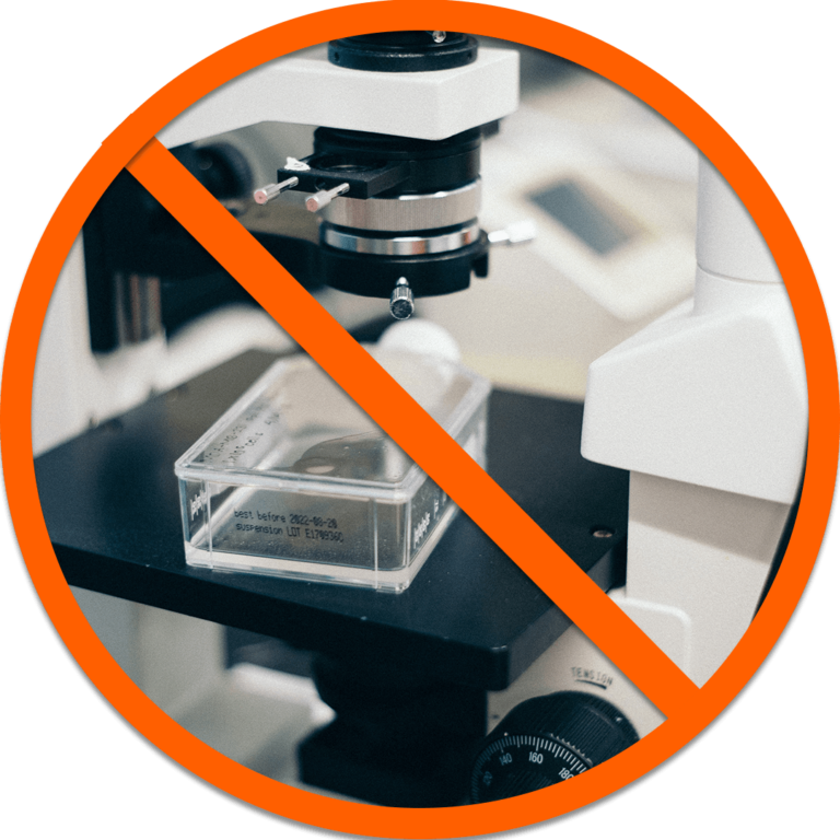
Having worked in the field of microscopy for over ten years, I know—from experience—that many microscope users lack the knowledge needed to avoid key errors and capture clear images for their research.
Take, for example, this microscope I recently fixed in our university lab.
Someone had loosened the stage of this microscope so much…that the ball bearings became loose and landed internally.
It’s a prime example of the kinds of uneducated mishaps that happen in labs.
And make no mistake…
A lack of microscopy education isn’t simply an inconvenience for organizations or universities. These knowledge gaps are also costly.
An uneducated mistake can cost a university thousands of dollars.
An uneducated mistake can cost researchers the chance of getting published.
An uneducated mistake can cost industry labs valuable time and resources.
For Example…
- A graduate student who lacks experience with a microscope may spend hours simply trying to learn the basic mechanics of his or her device…which keeps him or her from focusing on the research project at hand.
- A PhD candidate might choose the wrong objective for an experiment…leading to time wasted on needless troubleshooting, which also delays an important thesis deadline.
- A postdoc may neglect to save the raw images from an experiment. And—when the editors at a scholarly journal question his or her microscopy ethics—there’s no evidence to validate his or her time-consuming research. In turn, an important publication is lost.
- A new research scientist might skip an important step for permeabilizing a paraformaldehyde-fixated sample…causing the sample to lose its value for the experiment.
- A new researcher may damage a microscope objective worth 10s of thousands of dollars due to unfamiliarity of microscope operation.
As you can see, having a poor foundation in microscopy can keep you—or your organization—from quickly achieving key goals for your thesis, patent, or funding project.
In contrast, solid microscopy skills can accelerate how quickly you reach your objectives.
An Understanding Of Microscopy Principles Can Mean…
- The difference between an efficient lab or wasteful practices.
- The difference between a mediocre image or a stellar one.
- The difference between winning scholarly recognition or having top tier journals pass your research by.
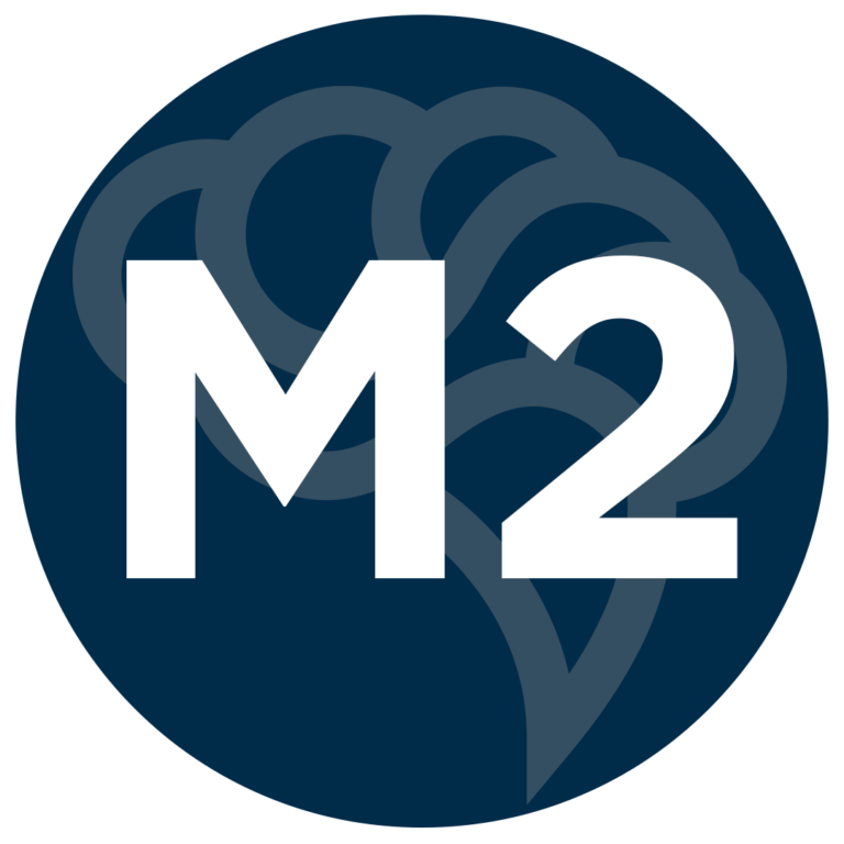
- Take a look at the images below to see what I’m talking about for yourself.
On the left, you’ll see an image before the right microscopy technique was applied.
On the right, you’ll see the same image. However, this image is taken after the proper microscopy technique has been executed.
The good news is, you no longer have to settle for images like those on the left.
You can give yourself or your students/research team a solid foundation in microscopy with…
The Exscope Private Group Provides Targeted Support
This Career Program’s Private Group Allows Scientists To Ask Microscopy Questions Freely
Whether you’re a graduate student or a core manager, the Expert Microscopy Career Program is here to replace embarrassed silence with helpful support.
The beauty of the Expert Microscopy Career Program is that scientists get instant access to a private group…
Where they can ask their questions freely.
Whether a scientist is having difficulty achieving a better resolution for an image or isn’t sure which fluorophore is best for his or her experiment, this exclusive group provides a safe forum to voice any challenges and enjoy sharing triumphs.
And there’s no need to worry whether others have access to this group.
Only fellow expert microscopy scientists and approved moderators will be able to see and respond to messages.
Below, you can get a glimpse into the kinds of interactions that take place in the Expert Microscopy Career Program group…
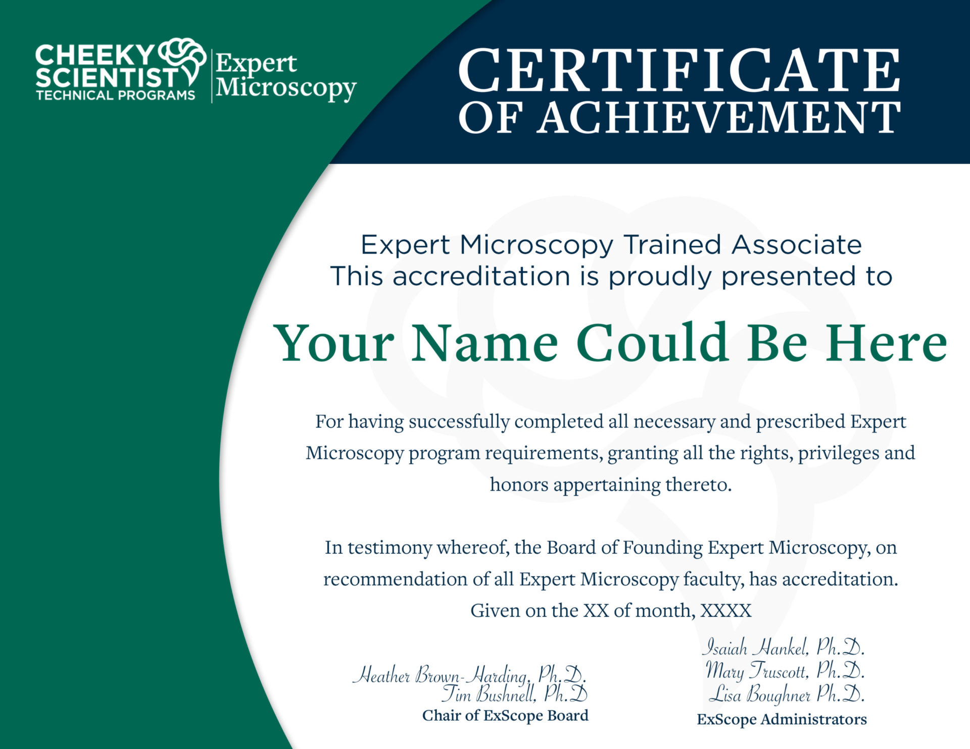
Those Who Complete The Expert Microscopy Career Program Can Expect An Official Validation From Our Board
Each member who completes the course materials will receive validation signifying that he or she is an expert microscopist. All Expert Microscopy Career Program scientists are welcome to leverage this validation to strengthen their resume and LinkedIn profile.
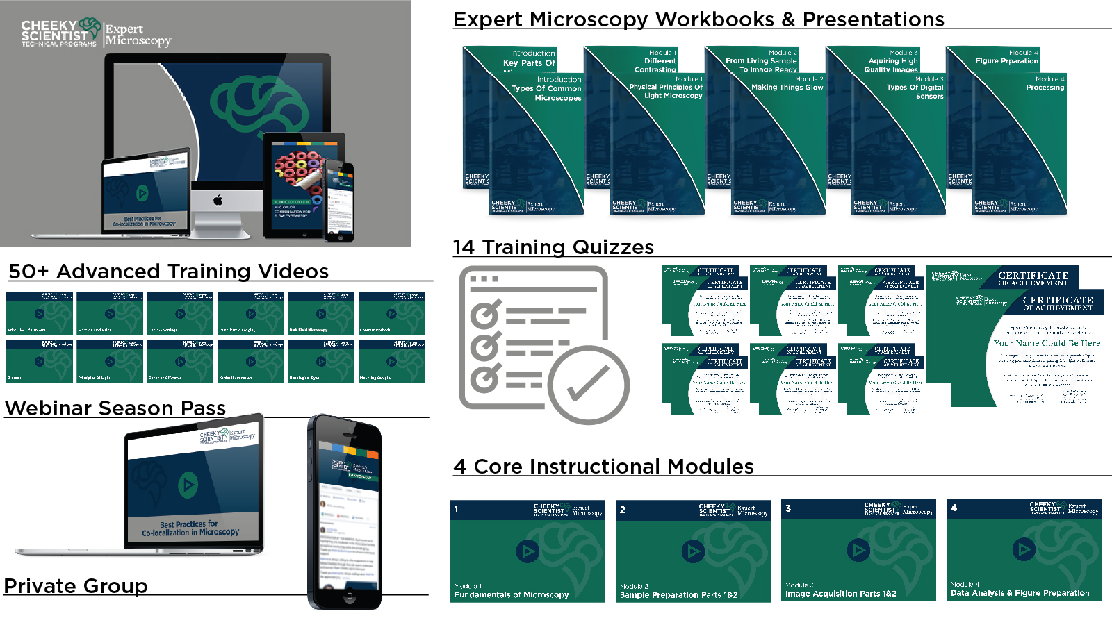
What’s Included In The Expert Microscopy Career Program?

Module #1: Principles of Light Microscopy Contrast Methods
The field of light microscopy is inseparable from the physical properties of light. This module will help students strengthen their understanding of this important concept.
Get ready to dive into…
- Important facts on the properties of light. Brush up on the physics behind microscopy. This module takes a look at the definition, properties, and behaviors of light.
- The relationship between a wavelength and microscope resolution. When it comes to microscopy, theoretical concepts can directly affect why an image looks different than expected. In this module, students will cover Airy discs, the Abbe diffraction limit, and the Rayleigh criterion…important factors for capturing high-quality images.
- The most important step in microscopy.Get the facts on illumination…as well as the six critical steps for achieving an ideal image for your research.
- Transmitted light methods. In microscopy, there are a number of methods that leverage transmitted light, and it’s important to have a broad knowledge of each technique. In this module, students learn a number of methods, including bright field, phase, differential interference contrast, Hoffman modulation contrast, polarization, and more.
Module #2: From Living Sample to Image Ready Fluorescence
Having cutting-edge microscope equipment doesn’t ensure you’ll capture high-quality images. The reality is, getting the best image for your experiment involves a number of factors that need to be carefully controlled. And preparing a sample is an important step for creating publication-worthy images. In this module, students dive into a number of important topics, including…
- Fixation methods you need to know. Before a researcher can capture an image, he or she needs to properly immobilize antigens while retaining cellular structure. This module helps students understand three techniques for fixating research samples…as well as important considerations for working with the various fixation methods.
- How to permeabilize a sample. Students will learn whether their fixation method will automatically permeabilize their samples…or if they need to use an additional method to ready a sample for imaging. Students will also discover the chemicals they can use to achieve this effect.
- Creating contrast for a sample. From histological dyes to indirect immunofluorescence, this module looks at three ways of creating contrast—giving students insight into some nuances for selecting a method.
- A scientific overview of the topic of fluorescence. Understand how this natural phenomenon works—from when a light photon hits an electron to the release of fluorescence. We’ll also cover the various filter options for a microscope as well as different sources of light.
- The pros and cons of different fluorophores. Get a better understanding of which fluorophores best suit a research project…for instance, which fluorophores can be genetically encoded or which can be toxic to the live cells in a sample.

Module #3: Image Acquisition Types of Detectors Acquiring High-Quality Images
Trial-and-error adjustments to a microscope are hardly a viable method for capturing a brilliant image for breakthrough research. In this module, students can strengthen their understanding of microscope cameras and key factors that influence image quality.
In this module, students can expect to learn…
- The basics of microscope cameras. The Expert Microscopy Career Program doesn’t assume students are experts in microscopic cameras. Instead, this module provides clear instruction on topics such as how pixilation affects image quality, when a high dynamic range is needed, and the difference between monochrome and color cameras…not to mention information on quantum efficiency and temporal resolution.
- What you need to know about photomultiplier tubes. Understand how these devices are different than cameras, how they work, and the quantum efficiency they provide.
- Important facts on image exposure, gain, binning, and more. If the settings of your microscope seem a mystery, this module will bring clarity to topics such as what range of exposure to use for fluorescent images and a pitfall to be aware of when it comes to digital offset.
- Facts on quantitative imaging and multidimensional acquisition. This module addresses intensity comparisons and size measurements…as well as how to leverage controls and a Z-stack for an image.

Module #4: Image Processing and Analysis Figure Preparation
Once a researcher has captured the perfect image of a sample, his or her work is not yet complete. In this module, students will get the facts on processing images and presenting an analysis that communicates the value of their research to journal editors and colleagues alike. Dive into topics such as…
- Why it’s critical to save your data. Understand the importance of backing up your files, and understand what is the industry-accepted software that microscopists use for processing.
- The different methods of processing microscopic images. In this module, students will discover when to use a Gamma adjustment…or when a flat-field correction can improve their images.
- Important components of image analysis. Discover how image segmentation, intensity measurements, co-localization, and high-throughput analysis can support your research objectives.
- Ethical considerations for image processing. Avoid an accusation of image manipulation by following best practices. According to the American Society for Microbiology, “as many as 35,000 papers in the literature are candidates for retraction due to inappropriate image duplication.” This module lays out three important principles students should follow as they prepare their images.
- How to ready your images for journal publication. Discover why Heather recommends Adobe Illustrator as your software of choice. This module also introduces students to considerations regarding their image resolution, format, color, and lookup tables.
- What you should know about graphing and statistics. Get an overview of how these concepts relate to microscopy research…and what details you should include in the methods section of your publications.
As Soon As You Join Exscope, You Will Also Get Immediate Access To The Following…
Understand the essentials of microscopy—from the parts of a microscope to preparing an image for a journal publication. Whether you need a resource to guide your university students or you’re a postdoc who needs better support, you’ll find our course doesn’t assume students have a strong background in microscopy—providing straightforward, easy-to-understand guidance.
- Lay a solid foundation for the rest of this course by understanding the basic mechanics of a microscope. The program starts by explaining the wide range of microscopes available and how they work…helping scientists know what to expect in a lab.
- Strengthen your understanding of the principles of light and how these affect microscopy activities. This program covers concepts such as Airy discs, illumination, and other essentials for helping students sharpen their skills in capturing high-quality images.
- Get the facts on how to prepare a sample for imaging and, ultimately, publication in a scholarly journal. Mentoring program members will receive important instruction for how to fix a sample, adjust microscopy settings, process an image, present an image to other scientists, and more.
Understand the nuances of capturing that perfect image. No matter how much effort a microscopist pours into a project, the quality of a sample can reflect the quality of his or her research—either positively or negatively. And the problem is, getting a crystal clear image isn’t as simple as following a 3-step process. Securing excellent images depends on a number of factors.
- Get information to decide which microscopy options are best for the needs of a sample. It’s important to realize that everything from the type of microscope used to the contrast technique chosen matters. You’ll find this course is helpful for making strategic microscopy decisions…before commencing research.
- Dive into the topic of fluorescence, taking a close look at this natural phenomenon and what materials are involved in an experiment—from filters to fluorophores.
Tap into the insights of an expert microscopist—who has experience in confocal microscopy, biological imaging techniques, and more. As mentioned previously, the Expert Microscopy Mentoring Program was developed by PhD Heather Brown-Harding.
- Let Heather’s workbooks guide you through important microscopy principles. There’s no need to worry about hard-to-understand language or convoluted descriptions. Heather’s expertise allows her to explain key microscopy concepts while keeping the course material accessible to a broad range of individuals.
- Further enrich the workbook material with videos featuring Heather, Dr. Isaiah Hankel and Dr. Tim Bushnell. Whether they’re covering information that reveals which microscope is suitable for the requirements of a research project or options for fixing a sample, these videos can help students stay engaged with the workbook material.
Gain a dashboard that hosts all the materials—including your workbooks, module videos, and more. With the Expert Microscopy Career Program, it only takes a simple login to access all your course resources for learning a new concept or brushing up on a lesson.
- Reinforce what you have learned with video transcripts and audio-only versions of the videos. The resources in this program accommodate different learning styles and the variety of situations students might encounter. Members can listen to MP4s while driving to the university or review their video transcripts while taking a lunch break.
- Leverage workbook exercises to test your newfound microscopy knowledge or to assess where your students/researchers are at in their learning journey. Each module contains some exercises to test comprehension of the course materials—as well as a module exam.
- Members who complete all course materials, will receive validation from the Expert Microscopy Career Program board. With this ribbon in hand, members can strengthen their LinkedIn profiles, add an additional qualification to their resumes, or assure a potential employer at their next job interview that they understand microscopy principles.
Gain access to the Expert Microscopy Career Program course, resources, and membership to the private online group for an entire year. There’s no need to worry that there’s only a few months to absorb the material presented in this online program. Instead—at their own pace—members can review this program over and over again until they’ve mastered the material.
- Share your challenges, chat with colleagues, and stay connected with fellow members in the private group. Members who’ve completed the course will still be able to interact with other members in the group or ask Heather advanced microscopy questions.
- Get advanced insights, thanks to continued webinars. This course includes a season pass to the Expert Microscopy Career Program’s webinars. From fluorescent proteins to live cell imaging, these sessions can deepen your understanding with additional education on advanced microscopy topics.
Frequently Asked Questions…
WHEN DOES THE EXPERT MICROSCOPY PROGRAM START?
Access to this career program begins as soon as you enroll. You’ll find that these courses are on-demand—so there’s no set pace or schedule you’re required to follow. Every student can choose the place, time, and setting of his or her education.
HOW DO I ACCESS THE COURSE MATERIALS?
Once you enroll in your career program, you’ll get a message from our team with your username and password. Use these credentials to sign into your dashboard and access all of your materials.
WHAT IF I MISSED AN ADVANCED CAREER WEBINARS?
No worries! We record each webinar, and your season pass gives you the ability to access these advanced sessions at a later date.
HOW LONG DO I GET ACCESS TO THE EXPERT MICROSCOPY CAREER PROGRAM?
Your membership to the Expert Microscopy Career Program lasts a full year. This gives you (or your students/researchers) plenty of time to access our materials and review them again and again.
WHAT ARE MY PAYMENT OPTIONS?
We offer two different payment options for this course. A 1 year membership or an extended membership that allows you to access the materials as long as you like.
Who Exscope Is NOT For…

It’s NOT For The Know-It-Alls
This is the graduate student who thinks he or she is an expert in microscopy. Or the core manager or lab manager who’s proud of his or her accomplishments—and expects his or her subordinates to attain to the same standard without providing support.
The problem is, a self-confident attitude will stunt academic learning. And a haughty attitude can stifle the intellectual curiosity of a research team.
The reality is, it’s easy for highly qualified individuals to specialize in a subset of science…without receiving important knowledge in microscopy.
So, if you’re a student or researcher who’s pretty confident in your microscopy knowledge…
Or if you’re a manager who expects your subordinates to get it right the first time…
Then the Expert Microscopy Career Program is simply not for you.
The Expert Microscopy Career Program can only help students who demonstrate intellectual humility or managers who believe in supporting the growth of their team.
It’s NOT For Those Who Dislike Applied Science
Passionate about reading scholarly journals…but hate venturing outside the theoretical realm to apply scientific concepts?
If this sounds like a description of your attitude…
Then the Expert Microscopy Career Program is probably not for you.
By definition, microscopy is a hands-on art. Microscopy involves understanding how a scientific phenomenon—such as fluorescence or interference—directly impacts a scientific experiment. It’s about executing practical solutions that work in harmony with scientific principles to capture that perfect image.
Microscopy isn’t for academics who dislike experiments or scholars who prefer to stay out of the lab.
The Expert Microscopy Career Program is about the hands-on application of science to help microscope users achieve faster, better experiments.

It’s Not For Scientists Who Are Okay With Irreproducible Experiments
This kind of person isn’t difficult to spot.
It’s the Phd who doesn’t mind getting a less-than-perfect image…so long as he or she can complete an assignment. Or the core manager who doesn’t mind cutting corners or watching students struggle…as long as he or she continues to enjoy a secure position at the university.
If this describes you, then you need to look for a different solution than the Expert Microscopy Career Program.
Our course is suited to help graduate students and researchers understand important principles of microscopy…so that they achieve their goals, streamline their experiments, and bring new scientific breakthroughs to light with incredible images.
If you’re willing to settle, then there’s no need to improve your skills—or those of your students—with this course. It simply isn’t for you.
Get Access to the Expert Microscopy Career Program & Network
If you’ve made the choice to improve critical microscopy knowledge with the Expert Microscopy Career Program…welcome!
Whether you’ve enrolled to educate your university students, bring a new research team member up to speed on best practices, or simply improve your own microscopy knowledge…
You’ll find your Expert Microscopy Career Program provides practical instruction in this important research skill.
Once you’ve enrolled, simply log into your dashboard and start educating yourself (or your students/subordinate researchers) with your step-by-step modules. And don’t forget to join the Expert Microscopy private group!
Tim Bushnell, PhD
Flow Cytometry Resource Facility Director at University of Rochester Founder, Expert Cytometry
Heather Brown-Harding, PhD
Assistant Directory of Microscopy at Wake Forest University Founder, Expert Microscopy

Swati Arora, PhD
Product Support Specialist at Zeiss Microscopy
Alex Woychex, PhD, MPH
Field Application Scientist At Akoya Biosciences
John-Michael Arpino, PhD
Advanced Imaging Specialist, Nikon Canada Inc.
Anil Shukla, PhD
Advanced Imaging Specialist, Nikon Instruments
We help PhDs from every discipline get into many different types of careers, including Expert Microscopy.
We show you how to leverage the skills you already have to get hired into a myriad of industry jobs, including Expert Microscopy.
We show you how to get the job, not how to do the job because each company has their own proprietary systems and will teach you how to do the job through on-the-job training.
But, before you can get this training, you have to get hired first.
That’s where we come in.
We will guide you on how to use your PhD to get hired into Expert Microscopy.