The Majority Of Flow Cytometry Experiments Are Not Reproducible – Your Experiments Can Be Different
Design Reproducible Flow Cytometry Experiments And Get Your Data Published, Funded & Patented
“The Expert Cytometry program was full of useful information and advice. The instructors were experienced, smart and funny. It was great to be able to go back through the instructional materials again and again.”
“It was very valuable and I would recommend that if your group is starting to use flow more heavily, that you bring Expert Cytometry in. There are nuances that I never knew and it immediately helped our group perform better experiments.”
“Medlmmune purchased a site license of Expert Cytometry. We have over 100 scientists using the program and the feedback has been phenomenal. I'm amazed at how much the scientists are coming to me with questions that are much more advanced than what I used to get before having access to ExCyte.”
Here’s A Look At Everything You Get As An Expert Cytometry Member...
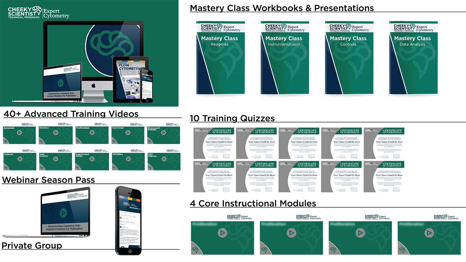
Each Expert Cytometry Core Instructional Module includes a one-hour instructional video along with a downloadable Expert Cytometry Workbook and Presentation. After the completion of each module, ExCyte’s members have the option to take a quiz. Administrators have the option to collect and record member quiz scores. There are 4 Core Instructional Modules, including:
- Instrumentation
- Reagents
- Controls & Compensation
- Data Analysis & Statistics
Once members have completed all 8 Module Quizzes, members can go on to access our Advanced Video series and test themselves with 6 Advanced Quizzes. Upon successful completion of all 14 Expert Cytometry Quizzes, members will be confirmed as esteemed flow cytometry professionals and receive a Expert Cytometry Validation.
Expert Cytometry Is For Scientists Who Want To:
- Stay Up To Date With Current Trends And Best Practices In Flow Cytometry.
- Gain Advanced Flow Cytometry Knowledge Quickly (Even If You’re A Beginner).
- Get Expert Information On Flow Cytometry Antibody Panel Design, Experimental Design, Data Analysis, And Statistics.
- Learn How To Control For And Compensate Any Type Of Flow Cytometry Experiment.
- Learn Specific Flow Cytometry Techniques And Methodologies.
Expert Cytometry’s Advanced Video Series Will Turn You Into A Flow Cytometry Expert
As part of the Expert Cytometry program, all members and administrators will receive access to Expert Cytometry’s Advanced Video series, which include over 10 one-hour instructional videos on:
- Advanced Statistics (Volume III)
- Advanced Experimental Design (Volume II)
- Advanced Proliferation
- Advanced Compensation (Volume IV)
- Advanced Data Analysis (Volume III)
- Advanced Controls (Volume IV)
- Advanced Antibody Panel Design (Volume II)
- Advanced Fluorescent Proteins
- Advanced Apoptosis
- Advanced FlowJo Analysis
- Advanced FACSDiva Analysis
- Advanced ImageStream Analysis & Many More
All Expert Cytometry Members Get Free Access To Our Bi-Monthly Webinars And Private Online Group
Members and administrators will also receive a one-year season pass to Expert Cytometry’s live webinar series and support through the Expert Cytometry Private Online Group.
This year’s mentoring series includes over 16 new webinars including:
- Microparticle Detection By Flow Cytometry
- Cell Sorting Rmax Methodology
- Fluorescent Protein Experiments By Flow Cytometry
- Macrophage And Monocyte Detection By Flow Cytometry
- Activated T-Cell Measurements By Flow Cytometry
- Advanced Instrumentation Comparison
- Advanced 10+ Color Methodologies ‘
- T Cell Biology By Flow Cytometry (Volumes 1 & II)
- B Cell Biology By Flow Cytometry (Volume 1 & II)
- Advanced Statistics (Volume V)
- Advanced Experimental Design (Volume IV)
- Advanced Proliferation (Volume II)
- Advanced Compensation (Volume VI)
- Advanced Data Analysis (Volume IV)
- Advanced Controls (Volume V)
- Advanced Antibody Panel Design (Volume IV)
- Advanced Apoptosis (Volume II)
- Advanced FlowJo SPICE Analysis (Volume II) & Many More
Are Your Flow Cytometry Experiments Reproducible?
It’s very likely that another lab could not reproduce your flow cytometry experiments.
Read this from Glenn Begley’s recent review in Circulation Research, entitled “Reproducibility in Science”.
“The estimates for irreproducibility based on these empirical observations range from 75% to 90%. These estimates fit remarkably well with estimates of 85% for the proportion of biomedical research that is wasted at-large. This irreproducibility is…seen across the spectrum of biomedical research.”
That’s right – 75% to 90% of biomedical experiments are not reproducible.
Government agencies are cracking down on reproducibility.
Read this from Andrew Bradbury’s review in Nature, entitled “Reproducibility: Standardize antibodies used in research”.
“Central to reproducibility in biomedical research is being able to use reagents that are identical to those described in publications. Alarmingly, there are serious flaws in the reliability of antibodies, the most widely used class of protein-binding reagent.”
If you want your data published, funded, and/or patented, you better start designing your experiments with reproducibility as your 1st priority.
You better start preparing credible and “approved” antibody panels too, no matter what kind of experiments you are running.
What Does This Mean For Flow Cytometry?
It means that your compensation methodology, including which exact antibody controls you use, their titrations, how you combined them, and so on must be in line with best practices.
The same holds true for your instrumentation settings.
And gating controls.
And statistical analyses.
The era of manually compensating your data and throwing together whatever antibodies you have in the lab to perform a flow cytometry experiment are over.
Getting guided properly in flow cytometry and keeping up-to-date with flow cytometry best practices is now required.
This Is Why We Created The Expert Cytometry Program
- ExCyte’s mission is simple – to ensure that your flow cytometry experiments are done properly and are reproducible.
- There is a lot of misinformation on how to properly set up and run flow cytometry experiments. As a result, many scientists are collecting misleading data and drawing incorrect conclusions.
- To that end, we developed ExCyte, which will give you the advanced flow cytometry mentoring you need to get your data published, funded, and patented.
- Expert Cytometry is the world’s only complete flow cytometry mentoring program for academic scientists and industry professionals.
- The Expert Cytometry program allows individual team members to work through mentoring modules at their own pace while completing individual quizzes after each module.
You Can Lose $500,000 Or More On Experiments That Are NOT Reproducible…
Recently we were engaged by a startup biotech to review some flow cytometry data that had been generated.
The company had very little experience in flow cytometry, but had purchased some promising intellectual property and results from early studies.
Based on that data, they engaged a CRO to generate more data to validate and extend the promising results.
An 8-color panel was designed and over 150 subjects were recruited.
Their blood samples were analyzed using this 8-color panel, and preliminary analysis by the CRO was performed.
In fact, they were in discussions with a second CRO to perform a second set of experiments to extend the results with the goal of marketing a blood test for a disease – even though the first cohort had not been reviewed.
This is where we came into the project – with over 1500 data files to analyze based on an 8-color panel designed by someone with minimal experience in flow cytometry.
Here Are Just Some Of The Problems We Found:
Panel Issues
- No viability dye
- Poor color choice (lots of overlap reducing sensitivity)
- No optimization of reagents (Titration/Voltage)
QC Issues
- No review of daily QC
- No method established to monitor voltages over time
- No Reference control
Control Issues
- No FMO controls for gating
- No reference control
- No saved compensation control data
- Use of isotype controls
Data Issues
- No plan for data analysis
- No idea of what populations were important
- No plan for downstream statistical analysis
- Not sufficient numbers of events collected for study
As We Delved Deeper Into The Data, We Determined That The Study Was Doomed Before It Even Started.
Over the course of our analysis, we had to exclude over 30 subjects for reasons that could have been resolved if proper flow cytometry practices had been observed.
For example, had there been proper compensation controls, another 10 samples could have been analyzed.
Issues like scatter parameters drifting were common in the data set, which suggested that no one was observing the flow cytometer as it was collecting data.
Furthermore, the QC reports showed that a laser had been changed out do to QC failures. Yet, this was not reflected in the voltages and 8 more samples were lost as data was off-scale due to this issue.
When all was said and done, ⅓ of the samples were excluded, dropping the power of the experiment dramatically.
In Conclusion, The Company’s Investors Lost $500,000, As Well As The Will To Redesign The Experiments Properly Or Do The Study Correctly.
The worse part was that a potential new diagnostic test that could have helped millions of people was lost too.
The only way to avoid these kind of negative scientific consequences is to get expert mentoring on proper experimental design and reproducibility.
Don’t wait until someone is digging through your data trying to figure out what went wrong.
Learn how to run your flow cytometry experiments correctly every time starting now.
Correct Experimental Design Is The Difference Between Success And Failure…
Steve had completed a PhD in T cell biology.
During his PhD, he had extensively used flow cytometry on an older, 2-3 color instrument.
Now, in his post-doctoral work, Steve was studying B-cells and using a digital 10-11 color flow cytometer.
His data and conclusions were going to form the basis of an NIH fellowship application.
The only problem was – Steve’s conclusions were wrong.
As we went through Steve’s data with him, we realized there were several fundamental flaws that made his interpretations incorrect.
This is what we figured out…
Steve didn’t understand how the multi-laser system worked. He thought that lasers turned on and off to excite the different fluorochromes.
This is not correct.
Steve also didn’t appreciate the best practices for compensation and gating controls. He relied on using outdated isotype controls and thought these controls were a perfectly acceptable way to identify positive cells from negative cells.
These controls are not acceptable.
As we tackled many different issues, Steve started quickly making changes to his protocols and, as a result, started generating much better data.
Soon after, Steve was awarded the aforementioned NIH fellowship and his flow cytometry data became central to the grants that his Principal Investigator successfully submitted.
Now, Steve is an NIH funded Assistant Professor on the
tenure track, with over a dozen publications, all of which rely heavily on flow cytometry.
If you don’t know how to set up, run, analyze, and report your flow cytometry experiment 100% correctly, you’re missing out.
You’re missing out on the grants, papers, and (most importantly) the thrill of correct discovery that could be yours.
Frequently Asked Questions
1. WHEN DOES EXCYTE START? HOW DO I ACCESS MY MATERIALS AFTER I SIGN UP? WHAT HAPPENS IF I MISS ONE OF THE NEW WEBINARS?
As a scientist, we know you’re busy. That’s why you get to determine the pace of the program. Once you sign up, you’ll receive a welcome email with your personal username and password. This login information will allow you to access to the private webpage where you can start to work your way through Module #1.
You can never “fall behind” in ExCyte because you have access to all of the Modules, Quizzes, and other materials right from the beginning. As a result, you can learn at your own convenience. All the videos and materials will be there for you for as long as you need them. All new webinars are recorded and archived so you can watch or re-watch them whenever you want.
2. YOU MENTIONED A EXCYTE PRIVATE GROUP—WHAT IS THIS AND HOW DO I GET THE MOST OUT OF IT?
There is a private and very friendly Facebook group of new and old Expert Cytometry members who are there to support you and each other with questions, answers, and comments on flow cytometry topics ranging from basic flow cytometry principles to cutting-edge methodologies.
In the past, getting high-level flow cytometry information and support meant waiting days, if not weeks, for help. Thanks to this group, you can get the help you need right away, often in the same day. Our founders, partners, and mentors are all in this group and willing to help you too.
3. WHAT ARE THE PAYMENT PLANS FOR EXCYTE? WHAT IF I DON’T LIKE EXCYTE? DO YOU HAVE A REFUND POLICY?
You can either pay $498 for a yearly subscription or pay $1,998 for a membership, where you will have all the updates of the ExCyte program without extra payment.
We are fully committed to helping you get the flow cytometry knowledge you need. If you’ve joined ExCyte, completed the materials, and still have doubts about the program, we’ll gladly refund 100% of your money up to two weeks (14 days) after you’ve joined.
Important: To be eligible for a refund, we require your completed answers to all ExCyte Quizzes and notes from all ExCyte Core Instructional Videos, Advanced Videos, Webinars, and all homework questions entered into the private group. This means you must take action in ExCyte.
We know that if you take action and do the ExCyte work, you’ll get results. If you do the work and don’t get value, then we will refund 100% of your enrollment fees. We do this because we’re looking for committed scientists only. We take our learning environment seriously and maintain a very high standard of action-oriented learning.
Hello Fellow Scientists,
I’m Dr. Tim Bushnell, the founder of Expert Cytometry. Our mission at Expert Cytometry is to make flow cytometry accessible, relevant, and enjoyable.
I’ve had a long history in the field of flow cytometry that started all the way back in graduate school.
Now, I’m the Director of the University of Rochester Medical Center’s Shared Resource Labs and a Council Member for the International Society for the Advancement of Cytometry (ISAC).
In today’s world, flow cytometry knowledge is robust.
Thanks to the Internet and the utility (and popularity) of this technology, flow cytometry knowledge is spread quickly, both far and wide.
However, are substantial problem has persisted in the field.
This problem is tied to a question every scientist should ask himself or herself on a daily basis…
Are You Collecting Misleading Flow Cytometry Data?
The biggest problem is there is a lot of misinformation on how to properly set up and run flow cytometry experiments. As a result, many scientists are collecting misleading data and drawing incorrect conclusions. This is why we created ExCyte.
ExCyte is the world’s only complete flow cytometry mentoring program for academic and industry professionals.
ExCyte allows individual team members to work through modules at their own pace while completing individual quizzes after each module. If you’re an organization with multiple ExCyte members, you can also appoint an administrator to support each member’s progress and additional members can be added at any time.
Here's What You Get Immediate Access To After Becoming An Expert Cytometry Member...

Module #1: Instrumentation
This module covers the process of generating data within a flow cytometer by covering the three major components of every instrument: fluidics, optics, and electronics.
We will explain how a particle goes from the “sample” to data file within the flow cytometer. The aim is to clarify how the three components of the cytometer are intertwined and work together to create the complex, rich data files that flow cytometry was developed to generate.

Module #2: Reagents
This module introduces users to the toolkit of specific probes most commonly used in flow cytometry to characterize the form and function of cellular and sub-cellular components and specimens.
We define fluorescence and describe common fluorescence processes that result in a measurable light signal that corresponds to biological phenotype or activity. Hundreds of natural and man-made fluorescent compounds are available to explore and measure biological phenomena. Different types of specific probes are introduced, including fluorescently tagged antibodies, transgenic fluorescent proteins, nucleic acid binding probes, fluorescent enzyme substrates, and cell division tracking dyes.
The merits of different fluorophores are compared in terms of brightness and usefulness in multiplex labeling strategies. Multicolor staining combinations maximize the amount of information gleaned from a single sample, but introduce special challenges due to overlap in the color of light emitted by many commonly used fluorophores. To separate and identify fluorescent signals from multiple probes we apply a mathematical correction to compensate for the natural crosstalk between many dyes.

Module #3: Controls
The aim of this module is to guide you, the user, regarding the possible sources of variation that may be encountered when preparing, collecting, and analyzing flow cytometry data.
Once the sources of variation are covered, techniques are presented to minimize variation and to yield high precision quantitation. Many of the techniques involve the use of appropriate bead standards for standardizing the instrument, compensation, flow rate, and event rate. In addition, tips are suggested regarding staining controls and the use of reference biological material. Lastly, data analysis strategies are presented that can be employed for objective region drawing.

Module #4: Data Analysis
In this module, you, the flow cytometry user, will receive an introduction to the basic parameters of flow cytometry data analysis.
Starting with the generation of the data file itself contained in the Flow Cytometry Standard, .FCS, we will discuss the various ways of presenting the raw data as graphical plots, how to draw regions and create gates, and the basic statistics used in many applications.
Lastly, we briefly touch on the subject of comparing data sets; that is, a group of control samples and a group of experimental samples, and how these data are generated.
We help PhDs from every discipline get into many different types of careers, including Flow Cytometry.
We show you how to leverage the skills you already have to get hired into a myriad of industry jobs, including Flow Cytometry.
We show you how to get the job, not how to do the job because each company has their own proprietary systems and will teach you how to do the job through on-the-job training.
But, before you can get this training, you have to get hired first.
That’s where we come in.
We will guide you on how to use your PhD to get hired into Flow Cytometry.